At Buxton & Bakewell Osteopathy Clinic, we help patients across Derbyshire and the Peak District find relief from trapped nerves and lower back pain. This blog helps explain everything you need to know.
The discs at the bottom of your lower back (L3/L4, L4/L5 and L5/S1) are the levels most likely to suffer from trapped nerves because these areas help support most of the weight of your upper body (two-thirds of your total body weight).

Trapped Nerves in the Back – Sciatica and Femoral Nerve Impingement
Our IDD Therapy programme is effective in treating trapped nerves, is non-invasive (unlike surgery) and is pain-free. IDD Therapy bridges the gap between what manual therapy cannot achieve and surgery. This therapy is the fastest growing therapy for trapped nerves and degenerative disc issues in the UK.
Each vertebra in the spine is given a number. In the lower back or ‘lumbar spine’, the vertebrae are numbered L1 to L5. The chest or ‘thoracic spine’ uses the letter T and is numbered T1-T12, and the neck or ‘cervical spine’ uses a C and is numbered C1-C7.
Slipped, herniated or disc bulges or protrusions usually occur at the bottom of your lower back at L3, L4 or L5 (and at C4/C5 or C5/C6 or C6/C7 at the bottom of the neck). There are also five rudimentary levels in the sacrum (although these are fused vertebra) where nerves exit, and these are numbered S1-S5.

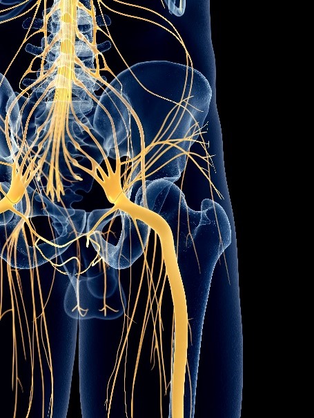
The discs at the bottom of your lower back (L3/L4, L4/L5 and L5/S1) are the levels most likely to suffer from trapped nerves because these areas help support most of the weight of your upper body (two thirds of your total body weight). The sciatic nerve runs from the bottom three vertebra (as seen below) and innervates the area around your hip, the back of the thigh and lower leg and foot.

If a nerve is trapped at L2 or L3 or L4 this will affect the femoral nerve (as seen below) and we suffer from femoral nerve impingement which provides both feeling and power to the front of the thigh. Therefore we experience pain in this specific anatomy.

These conditions cause a characteristic pain distribution down the leg. The areas of skin a single nerve innervates in the leg is called a dermatome. Each specific nerve will be responsible for sensory perception in a very specific area of skin (sensory perception being temperature, touch, vibration, pressure and pain). Therefore, if a nerve is impinged in the lower back, pain and pins and needles (or paraesthesia) will refer to any given dermatome.
So sciatic pain will potentially refer to any of those areas innervated from L3 to S3 levels (these levels innervate the back of the leg) and femoral nerve impingement will cause pain L2-L4 levels (these dermatomes innervate the front of the thigh) which provide both feeling and power to the front of the thigh.

Things to be aware of that are clinically significant and indicate that you need to take further action when you have sciatica are:
-
Severe impingement can weaken the ankle when walking (known as foot drop)
-
Progressive leg weakness
-
In extreme cases loss of bowel or bladder control and tingling/numbness in the groin area indicates a possible medical emergency.
For the femoral nerve, this generally provides both feeling and power to the front of the thigh (it innervates what we call the hip flexors and knee extensors). Movements such as climbing stairs (the knee may be unstable and prone to buckling) will be difficult as your thigh muscles will feel weak. Pain may also be felt on the side of the buttock, groin, inside of the knee and lower leg.
It is also worth mentioning that all the muscles in the legs are also innervated by nerves from different levels in the spine as well. These are called myotomes. The sciatic nerve, for example, will carry nerves for both sensory and motor innervation (motor as in ‘motor power’).
The information you give us in clinic and our clinical testing will help establish at which level in your spine you have a trapped nerve.
Causes of Trapped Nerves in the Spine
Herniated or Bulging Discs
There are a few terms commonly used when describing discs which we can quickly clarify.
A disc bulge is where the outer wall of the disc bulges out from its normal position. The disc wall is not broken, and the nucleus material is contained inside the disc. As the disc bulges, it may press against nerves directly. Often a bulge can be associated with a loss of disc height and this may lead to impingement of a nerve as it exits the spinal canal via a gap (called a foramen) between two vertebrae.

A herniated disc is the same as a prolapsed disc. This is where the nucleus of the disc breaks through the outer disc wall. There will be a loss of disc height as the disc loses pressure and the nucleus material can press directly on to the spinal nerves causing pain. Or, the material of the disc nucleus may act as a biochemical irritation to the nerve in which case the result is the same… pain!

A ‘slipped disc’ is an everyday expression which doesn’t have a true medical definition. It can imply a disc bulge or a herniation, usually a herniation.
This MRI below demonstrates a herniated disc pressing on nerves. The nerves are demonstrated by the broad white descending line seen in the scan. This is the spinal cord and departing spinal nerves. If you look carefully you can see the herniation making contact with these delicate structures.

The resulting pain from a herniated disc will often refer (hence the term radicular pain) down the pathway of a nerve and into the limb it innervates, causing either sciatica (in the case of the lower back) or pain into the neck, shoulder and arm (if in the neck). This can often be accompanied with pins and needles in the foot or hand depending on this location.
Our IDD Therapy programme is effective in treating trapped nerves, is non-invasive (unlike surgery) and is pain-free. IDD Therapy bridges the gap between what manual therapy cannot achieve and surgery. This therapy is the fastest growing therapy for trapped nerves and degenerative disc issues in the UK.
Trapped Nerves and Spinal Stenosis
Spinal stenosis refers to a build-up of bony deposits in the vertebrae. It is typically associated with the ageing process. As we get older, in the same way that our skin ages, so too do our discs. Everyone will have degenerative discs to some degree; it goes with the territory unfortunately.
In some cases, the loss of disc height as we lose water leads to more load pressure being exerted on the vertebrae.
The body reacts to the increased load by laying down more bone to reinforce the vertebrae. In some cases, the extra bone can narrow and exert pressure on the spinal cord (central stenosis) or exiting nerve roots (lateral stenosis).

Facet Joint Arthopathy
In the spine, facet joints link the vertebrae and are important for preventing excessive rotational and twisting forces which would damage the discs. They also share some of the load bearing of the spine.

When there is a loss of mobility in the spine, the facet joints bear a greater load than normal. This is particularly the case if there is some imbalance in the body and one side of the spine takes more strain than the other.
Imbalance in weight distribution not only adds to the stress borne by the facets but effectively deteriorates bone and cartilage. Constant movement on these worn structures activates an inflammatory reaction to the joint which is full of nerve endings. The result is chronic pain as the body continuously sends pain signals to the brain.
Just like the vertebra the body reacts to the increased load on the facet joints by laying down more bone in the joint margins (this is called facet joint arthropathy).

In some cases, the extra bone can narrow the gap where the nerves exit the spine and if the bone pinches against the bone, it can cause nerve root irritation (or a trapped nerve) causing lateral stenosis (or pressure on the nerve as it departs the spinal cord).
Degenerative Disc Disease
Disc degeneration is common in the neck (cervical spine) and lower back (lumbar spine). This is because these areas of the spine undergo the most movement and stress and are subsequently most susceptible to disc degeneration (as these bear much of our weight).
Degenerative disc disease refers to symptoms in the neck pain (this can refer to the shoulder) caused by wear-and-tear on a spinal disc. In some cases, degenerative disc disease also causes weakness, numbness, and hot, shooting pains in the arms and shoulder (radicular pain). Degenerative disc disease typically consists of a low-level chronic pain with intermittent episodes of more severe pain.
The discs are made of a compressible inner nucleus (nucleus pulposus) that deforms under load (as seen below) and an outer fibrous wall (anulus fibrosus) made of collagen.

The spongy intervertebral discs absorb shocks and pressure from the load of our bodies and squash as we lean or bend in any direction. They stop the vertebrae rubbing against each other (bone on bone) and they create a space between the vertebrae. This space is very important.
Each vertebra has a hole in the middle and when the vertebrae are stacked on top of one another combine to form a tunnel or canal through which the spinal cord travels down from the brain.
In children, the discs are about 85% water. The discs begin to naturally lose hydration during the ageing process. Some estimates have the disc’s water content typically falling to 70% by the age of 70, but in some people the disc can lose hydration much more quickly. Loss of hydration can be seen in the bottom two discs below.

As the disc loses hydration, it offers less cushioning and becomes more prone to cracks and tears. The disc is not able to truly repair itself because it does not have a direct blood supply. As such, a tear in the disc either will not heal or will develop weaker scar tissue that has potential to break again. At the same time, as the disc loses moisture and its structural integrity content it will protrude and bulge and can press on nearby nerves.
Our IDD Therapy programme is effective in treating trapped nerves, is non-invasive (unlike surgery) and is pain-free. IDD Therapy bridges the gap between what manual therapy cannot achieve and surgery. This therapy is the fastest growing therapy for trapped nerves and degenerative disc issues in the UK.


